Why the brain’s wires cross may trace back to ancient eyes
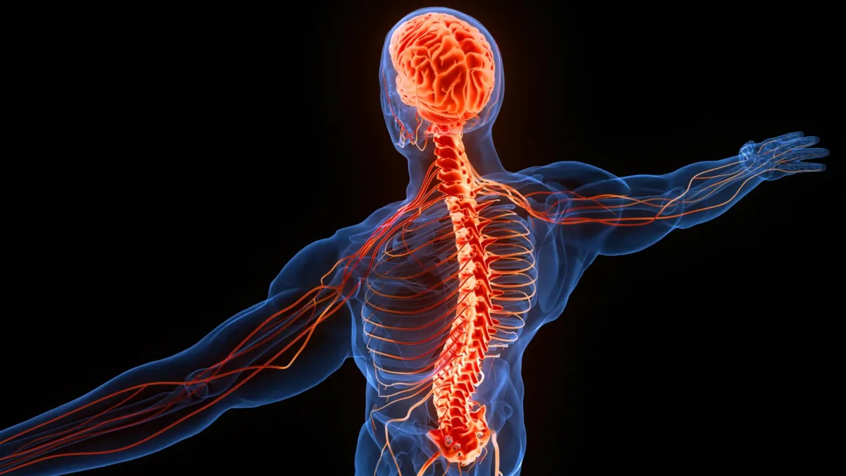
The left side of the brain controls the right side of the body, and the right side controls the left. Two researchers have proposed an explanation that reaches back hundreds of millions of years: evolution may have built new brain circuits around an ancient visual system.
Dr. Martin Wessendorf, a neuroscientist and associate professor emeritus at the University of Minnesota, studies how the brainstem regulates neurons in the spinal cord. Daniel Wang specializes in cognitive neuroscience and neurodegenerative diseases. Their study was published in Frontiers in Neuroanatomy.
The researchers examined several cross-wired neural circuits in four-limbed animals, meaning one side of the brain connects with the opposite side of the body. Their hypothesis suggests these connections may have developed because evolution built new circuits around an ancient visual system.
How the brain processes visual information
When light passes through the eye’s lens, the image projected onto the retina is reversed. Objects appearing to our right are projected onto the left side of the retina, and objects on our left onto the right.
Visual information travels along the optic nerve to the optic chiasm, where fibers are organized so that information from the right visual field reaches the left side of the brain, and information from the left visual field goes to the right side.
Some signals reach an ancient structure called the optic tectum, known in mammals as the superior colliculus. Located in the midbrain, it helps produce fast reactions to visual events, such as suddenly noticing something moving nearby. Each side contains a representation of the opposite visual field, and deeper within, information about physical sensations is also organized into corresponding maps.
Because the optic tectum exists in every vertebrate species, it is considered one of the most ancient structures in the vertebrate nervous system. Its early appearance may be especially important for understanding why so many other neural pathways cross sides.
Why sensory signals cross the brain and spinal cord
The researchers examined two major pathways that transmit sensations from the body to the brain. Both cross the center of the central nervous system and both appear to be widespread among tetrapods, the four-limbed animals including mammals, birds, reptiles and amphibians.
The first, the dorsal column pathway, carries information about gentle touch, vibration and the position of body parts. Signals from the limbs and torso travel upward through the dorsal portion of the spinal cord to another neuron in the brainstem, just above the spinal cord. From there, the pathway crosses to the opposite side. Fish lack this pathway, but scientists have identified it in amphibians, reptiles, birds and mammals. Its presence across these groups suggests the arrangement emerged early in tetrapod evolution and has remained largely conserved.
The second, the spinothalamic tract, transmits sensations involving pain, itching and certain types of touch. Unlike the dorsal column pathway, it crosses to the opposite side within the spinal cord itself. After crossing, signals travel upward to the thalamus, a brain region above the midbrain that plays a central role in processing sensory and movement information. This crossed arrangement is found in birds, mammals and reptiles. It also occurs in some cartilaginous fish, including sharks and rays, suggesting aspects of this organization have even deeper evolutionary roots.
How the brain controls movement across the body
The researchers identified three pathways involved in controlling skeletal muscles that also connect one side of the brain with the opposite side of the spinal cord.
The first is the tectospinal pathway, which helps the body respond rapidly to information from the environment. The optic tectum processes more than vision. It also receives information about sounds and physical sensations, organizing these signals into maps that align with its visual representation on the opposite side of the brain. The movement signals originate in the deepest layers of the optic tectum and travel through nerve fibers to the opposite side of the spinal cord. This pathway is present in most tetrapods and certain cartilaginous fish.
Another circuit is the rubrospinal pathway, thought to help animals control their limbs. It begins in a midbrain structure called the red nucleus and extends to the opposite side of the spinal cord. It is especially prominent in most mammals, although it has also been identified in other tetrapods and some cartilaginous fish.
The evolution of fine motor control in humans
The third pathway, the corticospinal tract, has a particularly important role in humans because it supports precise movements, including fine control of the fingers. Its evolutionary history is more complicated than that of the other pathways.
All vertebrates possess a telencephalon, the forward portion of the brain that accounts for much of the brain’s overall size in humans. However, only mammals have a cerebral cortex organized into six distinct layers. This six-layered cortex supports functions such as combining sensory information with memories, coordinating movement and processing language.
The corticospinal pathway extends from the cerebral cortex toward the spinal cord, but its function varies among animals. In most mammals, it primarily influences sensory processing. In primates, including humans, and certain other animals capable of highly precise movements, the pathway also reaches spinal cord regions responsible for controlling muscles, making intricate actions possible. Depending on the species, the tract can regulate sensory information or contribute directly to movement. In either case, its fibers cross the midline of the nervous system.
The researchers suggest humans may have developed advanced fine motor abilities by adapting an older neural circuit originally involved in controlling sensory input. Rather than creating an entirely new system, evolution may have given an existing pathway an additional purpose.
A 500-million-year-old explanation for the brain’s crossed wiring
The researchers propose that the earliest visual circuits helped establish a pattern in which sensory and movement pathways were organized across opposite sides of the nervous system. Later circuits may have inherited this arrangement as they evolved, particularly in tetrapods.